The Cell Cycle: Checkpoints, Mitosis, and the Logic of Division
How a cell decides to divide — and what happens when the brakes fail
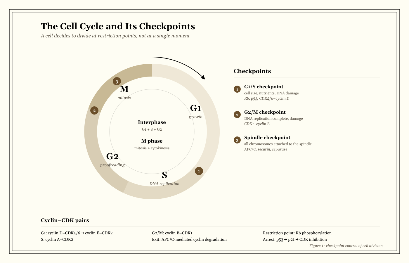
- Key Figure
- Leland Hartwell, Paul Nurse, and Tim Hunt (cell-cycle control)
- Field
- Twentieth century to present; Nobel Prize in Physiology or Medicine, 2001
- Method / Model
- Cell-cycle model; cyclin–CDK regulation and checkpoint control
- Core Problem
- How does a cell balance proliferation against genomic integrity, and what happens when the balance fails?
I. The Executive Dispatch (TL;DR)
- The cell cycle is a sequence of decisions, not just a loop. G1, S, G2, and M are coordinated by cyclin–CDK complexes and checkpoint surveillance.
- The restriction point is the point of no return. Once a cell passes the G1/S restriction point, it is committed to replicating its DNA and dividing, even if growth factors are later removed.
- Checkpoints protect genomic integrity. The G1/S, G2/M, and spindle checkpoints detect damage, incomplete replication, and unattached chromosomes.
- Cancer is a disease of deregulated control. Oncogene activation and tumour-suppressor loss combine to bypass checkpoints, drive proliferation, and increase genomic instability.
- The one-line exam takeaway: the cell cycle is controlled by positive drivers (cyclins/CDKs) and negative brakes (p53, Rb, CDK inhibitors), and cancer usually requires failures in both.
II. Historical & Intellectual Context (“The Backstory”)
From mitosis to molecular control
Nineteenth-century cytologists described mitosis as a visible process of chromosome movement. By the mid-twentieth century, cell biologists could measure the cell cycle and identify its phases. The molecular control of the cycle emerged from genetic studies in yeast and biochemical studies in sea urchin and frog eggs. Leland Hartwell identified cell-division-cycle (cdc) mutants in budding yeast; Paul Nurse used fission yeast to identify CDK1 (cdc2); Tim Hunt discovered cyclins in sea urchin eggs. They shared the 2001 Nobel Prize in Physiology or Medicine.
The result was a model of the cell cycle as a regulatory network: cyclins rise and fall, CDKs phosphorylate targets, and checkpoints integrate signals. The model was soon connected to cancer biology. Oncogenes and tumour suppressors were found to be components of the same machinery, and the cell cycle became the central framework for understanding uncontrolled proliferation.
Why the cell cycle matters
The cell cycle is the basis of growth, development, tissue repair, and reproduction. It is also the target of many cancer therapies. Drugs that inhibit CDKs, microtubules, or DNA replication exploit the differences between dividing and non-dividing cells. Understanding the cycle is therefore both a core exam topic and a practical problem in medicine.
III. Structural & Formal Architecture
The phases
The cell cycle has two major parts: interphase and M phase.
| Phase | What happens | Key control |
|---|---|---|
| G1 | Cell growth; assessment of nutrients, size, and DNA damage | Restriction point; Rb, p53, CDK4/6–cyclin D |
| S | DNA replication; sister chromatids form | CDK2–cyclin E/A; licensing and origin firing |
| G2 | Further growth; repair and preparation for mitosis | G2/M checkpoint; CDK1–cyclin B |
| M | Mitosis and cytokinesis | Spindle checkpoint; APC/C |
The duration of each phase varies by cell type. A typical cycling mammalian cell might spend roughly 10–12 hours in G1, 6–8 hours in S, 3–4 hours in G2, and 1 hour in M, but these values are illustrative, not universal.
Cyclin–CDK control
Cyclin-dependent kinases are the engines of the cell cycle. They are inactive alone and require a cyclin partner for full activity. Different cyclin–CDK pairs drive different transitions:
- G1: cyclin D–CDK4/6 phosphorylates Rb, releasing E2F and allowing S-phase genes to be transcribed.
- G1/S: cyclin E–CDK2 drives the G1/S transition and origin licensing.
- S: cyclin A–CDK2 supports DNA replication.
- G2/M: cyclin B–CDK1 (also called MPF) drives entry into mitosis.
- Exit: APC/C ubiquitinates cyclins, targeting them for degradation and allowing mitosis to finish.
The system is not a simple linear cascade. Positive feedback loops, phosphorylation switches, and degradation create irreversible transitions. That is why the cell cycle can be described as a series of bistable switches rather than a smooth clock.
Checkpoints
Checkpoints are surveillance mechanisms that delay progression until conditions are met. They are not permanent stops; they are decision points.
- G1/S checkpoint (restriction point): monitors cell size, nutrients, growth factors, and DNA damage. Rb and p53 are central.
- G2/M checkpoint: monitors DNA replication completion and damage. CDK1–cyclin B is inhibited if repair is needed.
- Spindle checkpoint (M checkpoint): monitors chromosome attachment to the mitotic spindle. The APC/C is inhibited until all chromosomes are properly attached.
IV. The Chronicle: Process and Mechanism
Step 1 — G1 and the restriction point
The Action. The cell grows and responds to growth factors. If conditions are favourable and DNA is intact, it passes the restriction point and commits to division.
Key Evidence. Rb is hypophosphorylated in quiescent cells and binds E2F, repressing S-phase genes. Cyclin D–CDK4/6 phosphorylates Rb, releasing E2F and allowing transcription of cyclin E and other S-phase genes. p53 can induce p21, a CDK inhibitor, in response to damage.
The Subtext. The restriction point is the cell’s commitment decision. Before it, the cell can return to G0; after it, the cell is committed to completing the cycle, even if growth factors are removed.
Step 2 — S phase and DNA replication
The Action. DNA replicates, and each chromosome becomes two sister chromatids. Replication origins fire in a controlled order, and each origin fires only once per cycle.
Key Evidence. Licensing factors (MCM complexes) are loaded in G1 and activated in S phase. CDK activity prevents re-licensing, ensuring that the genome is replicated exactly once.
The Subtext. The once-per-cycle rule is essential for genomic stability. Failure leads to re-replication, DNA damage, and aneuploidy.
Step 3 — G2 and the G2/M checkpoint
The Action. The cell checks for DNA damage and incomplete replication. If damage is present, the cycle is delayed while repair occurs. If repair fails, the cell may undergo apoptosis.
Key Evidence. CDK1–cyclin B is kept inactive by Wee1 and Myt1 kinases and by inhibitory phosphorylation. Cdc25 phosphatases remove the inhibitory phosphate to activate CDK1 and drive mitosis.
The Subtext. The G2/M checkpoint is a quality-control step before the cell commits to chromosome segregation. It integrates damage signals with the mitotic machinery.
Step 4 — Mitosis
The Action. The cell divides its nucleus through prophase, prometaphase, metaphase, anaphase, and telophase.
| Stage | What happens |
|---|---|
| Prophase | Chromosomes condense; centrosomes separate; spindle begins to form |
| Prometaphase | Nuclear envelope breaks down; kinetochores attach to microtubules |
| Metaphase | Chromosomes align at the metaphase plate; spindle checkpoint monitors attachment |
| Anaphase | Sister chromatids separate; APC/C triggers securin degradation and separase activation |
| Telophase | Nuclear envelopes reform; chromosomes decondense |
Key Evidence. The spindle checkpoint inhibits APC/C until all kinetochores are attached. Once satisfied, APC/C ubiquitinates securin, releasing separase, which cleaves cohesin and allows sister chromatids to separate.
The Subtext. Mitosis is a mechanical process with a surveillance system. The checkpoint couples chromosome attachment to the irreversible step of anaphase.
Step 5 — Cytokinesis and exit
The Action. The cytoplasm divides. In animal cells, an actomyosin contractile ring pinches the cell into two daughters. In plant cells, a cell plate forms.
Key Evidence. RhoA and formin drive actin assembly; myosin II generates contractile force. The midbody forms between daughter cells and is involved in abscission.
The Subtext. Cytokinesis is distinct from mitosis. A cell can complete nuclear division without cytoplasmic division, producing a multinucleate cell.
Step 6 — Quiescence, senescence, and death
The Action. Not every cell divides. Cells can exit the cycle into G0 (quiescence), enter senescence (permanent arrest), or undergo apoptosis.
Key Evidence. G0 cells may re-enter the cycle upon stimulation; senescent cells remain metabolically active but do not divide; apoptotic cells activate caspases and are cleared.
The Subtext. The default state of many cells is not division. Proliferation is a controlled exception, not the rule.
V. The Editorial Board: Model Limits, Assumptions, and Debates
The cell cycle is not a universal clock. The classic model is based on a few cell types, especially yeast and cultured mammalian cells. Embryonic cells, stem cells, and some differentiated cells have modified cycles with shortened gaps or specialized controls. The model is a framework, not a universal timetable.
Checkpoints are not absolute. A checkpoint delays progression; it does not guarantee repair. If damage is irreparable, the cell may undergo apoptosis or enter senescence. Cancer cells often bypass checkpoints rather than deleting them entirely.
p53 is a tumour suppressor, not an oncogene. This is the most common exam error. p53 loss or mutation removes a brake; oncogenes such as RAS or MYC are drivers that promote proliferation when activated. Cancer typically involves both.
Aneuploidy and genomic instability. Failures in chromosome segregation, centrosome duplication, or cytokinesis can produce aneuploid daughter cells. Aneuploidy is common in cancer but is also found in normal tissues, so it is not sufficient by itself to explain malignancy.
Therapeutic targeting. CDK4/6 inhibitors (e.g., palbociclib) exploit the G1/S transition in hormone-receptor-positive breast cancer. Taxanes stabilize microtubules and activate the spindle checkpoint. Understanding the mechanism explains the side effects and resistance patterns.
Reproducibility and measurement. Cell-cycle analysis depends on the method: flow cytometry of DNA content, BrdU/EdU incorporation, phospho-histone H3, and live-cell imaging measure different things. A “G1 arrest” by DNA content may include G0 cells; a “mitotic index” depends on the marker used.
VI. Exam & Essay Sparkplugs
1. “The restriction point is the most important decision in the cell cycle.” Discuss.
Winning Thesis Blueprint. The restriction point commits a cell to division and integrates growth, nutrient, and damage signals, making it the key regulatory gate. However, later checkpoints can still arrest or kill the cell, so commitment is not the same as completion.
Supporting points.
- Rb phosphorylation and E2F release.
- p53/p21-mediated arrest after damage.
- The distinction between commitment and completion.
2. “Cancer is a disease of checkpoint failure.” How far do you agree?
Winning Thesis Blueprint. Cancer involves bypass of checkpoints, but it also requires proliferative drive, evasion of apoptosis, and genomic instability. Checkpoint failure is central, but it is not the whole story.
Supporting points.
- Oncogene activation and tumour-suppressor loss.
- p53 and Rb pathways.
- Aneuploidy and clonal evolution.
3. “Mitosis and cytokinesis are separate processes.” Explain why the distinction matters.
Winning Thesis Blueprint. Mitosis divides the nucleus; cytokinesis divides the cytoplasm. The distinction matters because failures in either process produce different outcomes: binucleation, aneuploidy, or failed cell separation.
Supporting points.
- The stages of mitosis and the spindle checkpoint.
- The actomyosin ring and cell plate.
- Examples of cytokinesis failure and multinucleation.